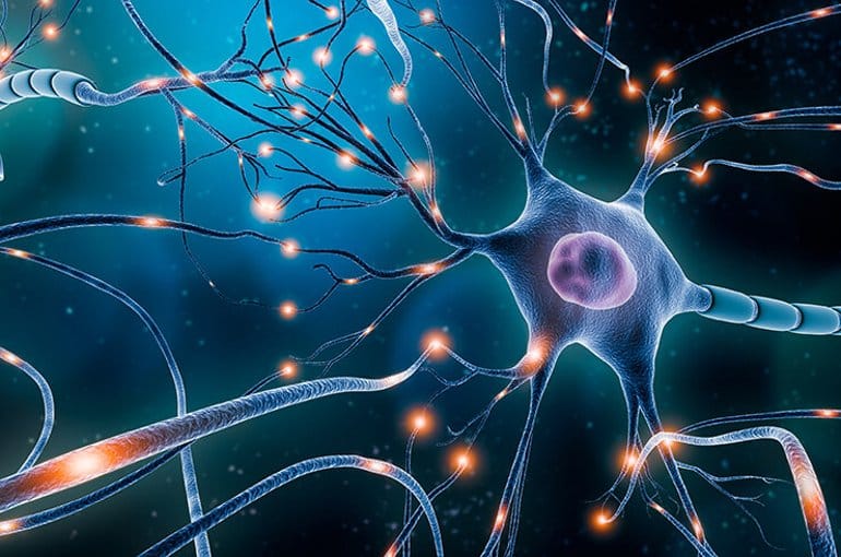
Neurons
The functioning of the brain is based on several billion nerve cells called “neurons” that receive and send electrochemical signals or “messages”. Neurons are the functional unit of the brain, made up of a cell body, dendrites, and an axon. The axon sends electrochemical signals to other neurons and the many small hair-like dendrites receive the signals. Signals along the axon are protected by a layer of myelin. This protective sheath makes sure the messages are sent quickly from one end of the axon to another.
When the signal reaches the end of the axon, chemicals called neurotransmitters are released from synaptic vesicles and are picked up by the dendrites of the next neuron. These can then go on to generate another electrical signal, called an action potential. The connections between neurons are called synaptic connections and during the first few years of life, trillions of them are formed. The sheer number of these connections are what allows children to understand & process all the information they receive.
An enriched environment can lead to more & stronger synaptic connections.
Neural Pathways
When children are born, their brains are a mass of neurons, ready to be wired or programmed through use and experience. Some hardwiring is already present to regulate breathing, reflexes, body temperature, and heartbeat. Billions of other neurons are ready to be connected, but they must be used for connections to be made. Unused neurons do not survive.
This discarding of synaptic connections (“pruning”) eliminates those that are not used in favor of those that are formed and used through everyday experiences. The early experiences of children play a critical role in determining which neurons are to be used and which are to die. As the child grows, a complex system of synapses and neural pathways is formed. The pathways that are repeatedly activated or used are protected and retained into adulthood.
The wiring of the brain occurs very rapidly during early childhood:
- By age 1, scans of the brain are more similar to an adult brain than a newborn.
- By age 2, the number of synaptic connections reach adult levels.
- By age 3, a child’s brain has about 1 quadrillion (1,000 trillion) connections, twice the number of an adult’s brain, and is 2 ½ times more active.
- By late adolescence, about half of the synaptic connections have been discarded.
As a child interacts with the environment, various stimuli enter the body via the five primary senses – sight, smell, sound, taste and touch. These experiences are then converted into electrical/chemical messages that travel through the spinal cord to the thalamus – a structure in the center of the brain. This important structure sends the incoming stimuli to one of the four lobes (occipital, temporal, parietal and frontal) of the brain for further processing.
Young children’s brains are growing at an incredible rate. By the time a child reaches the age of 5, more than 100 billion neurons have made connections within the cerebral cortex. Many of these neurons, if not used, die. The stronger the connections between neurons, the stronger and faster the reaction will be in recalling information.
Sections of the Brain
Here's a quick peek at the different sections of the human brain.
The Frontal Lobe is the largest part of the cortex, with the most complex functions, including sensorimotor processing and cognition. It includes the Pre-Frontal Cortex which processes “higher” brain functions, including our ability to plan, reason, make judgments, dream, and regulate emotional impulses.
The Occipital Lobe houses the primary centers for processing visual stimuli. Incoming stimuli are first processed and organized in the primary visual perception area. It then travels to the visual association area, where it is compared to what the brain has seen before.
The Temporal Lobe houses the primary center for processing auditory stimuli. It also has 2 primary areas, one for processing & organizing and the other for making associations. In coordination with the hippocampus, it is essential for long-term memory functioning.
The Parietal Lobe analyzes and integrates somatosensory stimuli from different parts of the body, to provide a sense of spatial awareness and maintain focus or spatial attention.
The Motor Cortex generates the neural impulses controlling the execution of movement.
The Thalamus is the information messenger between the cortex, brain stem, and other structures.